Massive 18.5 kg fibroid removed from 56-year-old woman, experts highlight need for early diagnosis
For nearly six years, a 56-year-old woman carried a secret burden — a growing weight in her abdomen that slowed her down, made walking painful, and left her breathless with even small movements. What she thought was “just bloating” turned out to be one of the largest fibroid uteruses ever removed in recent years, weighing a staggering 18.5 kilos.
The life-changing surgery was performed at BLK-Max Super Speciality Hospital by a multidisciplinary team led by Dr Dinesh Kansal, Head of Obstetrics & Gynaecology and Laparoscopic Surgery. “This was far more challenging than her scans had suggested,” Dr Kansal said. “The benign tumour had severely distorted her anatomy, compressing the bladder, intestine, and ureter. Scar tissue from earlier surgeries made it even more complex.”
The multi-hour procedure required detailed pre-operative planning and real-time decisions in the operating theatre. The patient was discharged just four days later and has since returned to an active lifestyle.
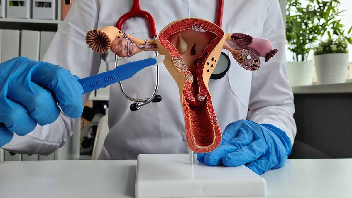
What are fibroids?
Fibroids are non-cancerous growths in the uterus and are common among women of reproductive age, but can go unnoticed until they cause severe discomfort. Doctors warn that delaying medical evaluation can turn treatable conditions into surgical challenges.
“If she had sought help earlier, her surgery could have been much simpler,” Kansal said. “This case is a reminder to take persistent symptoms seriously.”
Commonly called Leiomyomas, or fibroids, benign uterine tumors most commonly develop and grow as a result of imbalances in the hormones, particularly estrogen and progesterone. As per a paper published in the Journal of Pathology and Translational Medicine, gigantic benign tumors can be managed complication-free with proper diagnosis and surgical expertise.
In one of the earlier such cases at Motherhood hospital in Pune, investigations revealed two ovarian cysts — one measuring 5x6 cm on the right ovary and another measuring 3x4 cm on the left ovary - in a 29-year-old woman. Additionally, parts of her intestine were adhered to the cysts, a condition known as peritoneal endometriosis, where the endometrial lining extends outside the uterus and implants on the peritoneum — the thin layer of tissue lining the abdominal cavity and covering the abdominal organs. The space behind the uterus, called the Pouch of Douglas (POD), a common site for endometriosis growth, was also blocked. Dr. Sushil D. Garud, Consultant - Obstetrics, Gynecology, and Laparoscopic Surgery at the hospital, performed a laparoscopic surgery that lasted for nearly two hours, during which he successfully removed the ovarian cysts and endometrial implants from the woman's abdomen.
Health